
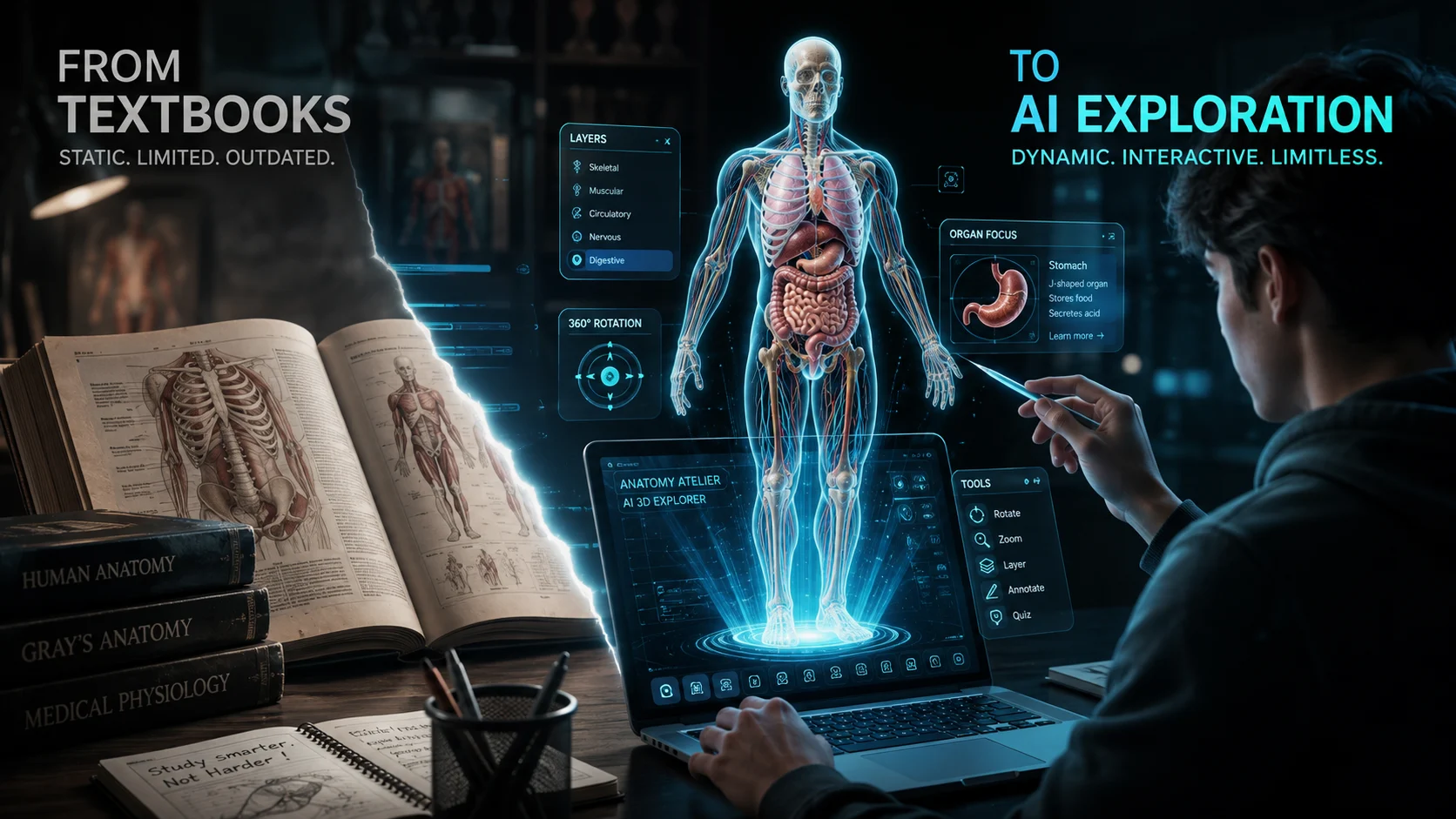
Static medical textbooks just suffered a decisive blow as AI-powered 3D learning takes over. Studies show 80% of medical students struggle with spatial memorization from 2D diagrams, while interactive 3D tools increase retention by up to 40% and reduce study time by 30%.
Why are classrooms still relying on heavy printed atlases when AI can build depth instantly? This shift has transformed medical education and accelerated how future doctors master human anatomy.
Why Anatomy Atelier Stands Out
What makes this tool different from traditional learning platforms?
Anatomy Atelier turns complex bodily systems into fully rotatable, interactive models. Students can zoom, isolate, and dissect anatomical layers directly from a web browser or mobile tablet.
- Interactive Medical Learning: Manipulate organs, nerves, and muscle groups in 360-degree space.
- Instant Layer Isolation: Hide superficial tissue to examine deep vascular structures underneath.
- Next-Gen Anatomy Explorer: Explore organ cross-sections without physical cadaver lab constraints.
The AI Tools Behind the 3D Models
How does AI generate hyper-accurate 3D anatomical structures?
By utilizing text-to-3D pipelines, automated mesh refinement, and volumetric segmentation, the platform builds high-resolution meshes in seconds rather than weeks.
| Feature | Traditional 3D Modeling | AI-Assisted Modeling |
|---|---|---|
| Development Time | Weeks per organ model | Seconds using automated pipelines |
| Anatomical Precision | Manual adjustments required | Derived directly from volumetric scans |
| Workflow Scalability | High cost, slow iteration | Faster iteration and instant updates |
| Interactivity Level | Fixed pre-rendered angles | Full real-time rotation and scaling |
And the production time keeps shrinking — raising a critical question about future medical curricula.
What Medical Students Gain
When students can manipulate models in real-time, spatial relationships become immediately clear. Complex neuroanatomy that took weeks to memorize can now be visualized in minutes.
- Faster Learning: Accelerates comprehension of complex anatomical layers.
- Better Spatial Recall: Builds an intuitive mental map of human systems.
- High Accessibility: Eliminates the need for expensive cadaver labs for basic revision.
“The real value isn’t just better graphics — it’s giving students a faster, more intuitive mental model of complex anatomical structures.” — Digital Learning Specialist
Why Textbooks Are Losing Ground
Could flat diagrams make traditional memorization obsolete?
Printed atlases offer static cross-sections that force students to imagine depth manually. AI-generated models remove this cognitive barrier by rendering high-fidelity depth instantly.
- No Static Limits: Pan around structures rather than guessing hidden angles.
- Dynamic Dissection: Toggle visibility on surrounding organs to isolate specific pathways.
- Portable Studying: Carry a complete virtual cadaver lab inside a lightweight tablet.
And the biggest shift may not be technical — it may be psychological.
Only if education could be this interactive ❤️🔥
— The Bugged Dev (@thebuggeddev) August 2, 2026
I’ve had a looong wish to build something genuinely useful through vibe coding, and I finally did it.
A 3D human anatomy application built with @threejs using GPT 5.6 Sol.
It all started with a single design image that I created… pic.twitter.com/vuevYA24Sn
The Future of Anatomy Training
Will universities soon replace physical atlases entirely?
As generative AI pipelines continue to improve, integration with extended reality (XR) headsets and AR/VR classrooms will accelerate. Medical schools are already evaluating how AI-built 3D models can standardize training across hospitals globally.
Official Portals & Research
Platform & Medical Research: • Anatomy Atelier Official Platform • National Center for Biotechnology Information (NCBI) Research • Medgadget Digital Health News • MobiHealthNews Innovation Coverage